MohsPedia
Comprehensive encyclopedia of Mohs surgery techniques, flaps, grafts, sutures, anatomy, and regional reconstruction.
Anatomy Atlas
Interactive anatomical reference: 18 face zones & 10 body regions with danger zones and reconstruction principles.
DermSurgery 101
Foundational curriculum for dermatologic surgery: sutures, flaps, grafts, and essential surgical techniques.
Journal Club
165 curated articles from the Northwestern Medicine Dermatologic Surgery Journal Club (2022–2026).
35 articles available
Flaps
Local tissue rearrangement. Advancement, rotation, transposition, and interpolation flaps.
Advancement Flaps
Advancement flaps move tissue in a single linear vector toward the defect without rotation or lateral movement. They are among the most commonly used closures in Mohs reconstruction, particularly on the forehead, scalp, and upper lip. Variants include U-plasty, H-plasty, T-plasty, L-plasty, island pedicle (V-to-Y), and crescentic advancement.
Rotation Flaps
Rotation flaps pivot tissue about a fixed point along a curvilinear arc to fill an adjacent triangular or circular defect. The arc length, pivot point mechanics, and back-cut placement are the critical design variables. Common variants include the standard rotation flap, O-to-Z bilateral rotation, and the dorsal nasal (Rieger) rotation flap.
Transposition Flaps
Transposition flaps are lifted and moved over intervening normal tissue to reach the surgical defect. They redirect wound closure tension and can recruit tissue from areas of greater laxity. Key variants include the Z-plasty, rhombic (Limberg) flap, bilobed flap, nasolabial transposition, and banner flap.
Interpolation Flaps
Interpolation (importation) flaps are two-stage pedicled flaps in which the flap base is not immediately adjacent to the defect. The pedicle bridges over or under intervening tissue, requiring a second-stage procedure (pedicle division) after the flap has established neovascularization at the recipient site. Major types include the paramedian forehead flap, melolabial interpolation flap, retroauricular flap, and Abbe (lip switch) flap.
Island Pedicle Flaps (V-to-Y Advancement)
Island pedicle flaps (V-to-Y advancement flaps) derive their blood supply from a deep subcutaneous pedicle, making them unique among local flaps. The skin island is completely circumscribed and advanced into the defect while remaining tethered only to the underlying subcutaneous tissue. This category includes the classic V-to-Y island pedicle and the modified single-sling myocutaneous island pedicle flap described by Willey et al.
Grafts
Full-thickness, split-thickness, and composite tissue transfer techniques.
Full-Thickness Skin Grafts (FTSG)
Full-thickness skin grafts harvest the complete epidermis and dermis, providing the best color and texture match with the lowest contraction rate among graft types. They are the most commonly used graft in dermatologic surgery, particularly after Mohs micrographic surgery for facial defects.
Composite Grafts, Split-Thickness Grafts, and Free Cartilage Grafts
Composite grafts transfer multiple tissue layers (skin, cartilage, and sometimes mucosa) as a single unit, primarily for nasal alar and auricular reconstruction. Split-thickness skin grafts (STSG) harvest epidermis with variable dermis thickness and are preferred for large or poorly vascularized wound beds. Free cartilage grafts provide structural support without skin coverage.
Sutures & Closures
Suture materials, needle selection, and closure techniques for optimal outcomes.
Suture Materials
A thorough guide to suture materials used in dermatologic surgery, covering physical properties, absorbable vs. nonabsorbable classifications, natural vs. synthetic materials, and needle anatomy. Understanding suture characteristics is fundamental to selecting the appropriate material for each clinical scenario.
Suturing Techniques
A thorough guide to suturing techniques in dermatologic surgery, from the workhorse buried vertical mattress to superficial closure methods. Covers suture placement biomechanics, tissue eversion principles, tension management, and suture removal timing.
Surgical Technique
Mohs micrographic surgery principles, tissue processing, and margin evaluation.
Mohs Micrographic Surgery Technique
Mohs micrographic surgery (MMS) is a specialized surgical technique that provides 360-degree peripheral and deep margin assessment, evaluating approximately 100% of the surgical margin. The Mohs surgeon serves as both surgeon and pathologist, performing staged excision with complete margin evaluation to achieve the highest cure rates while maximizing tissue conservation.
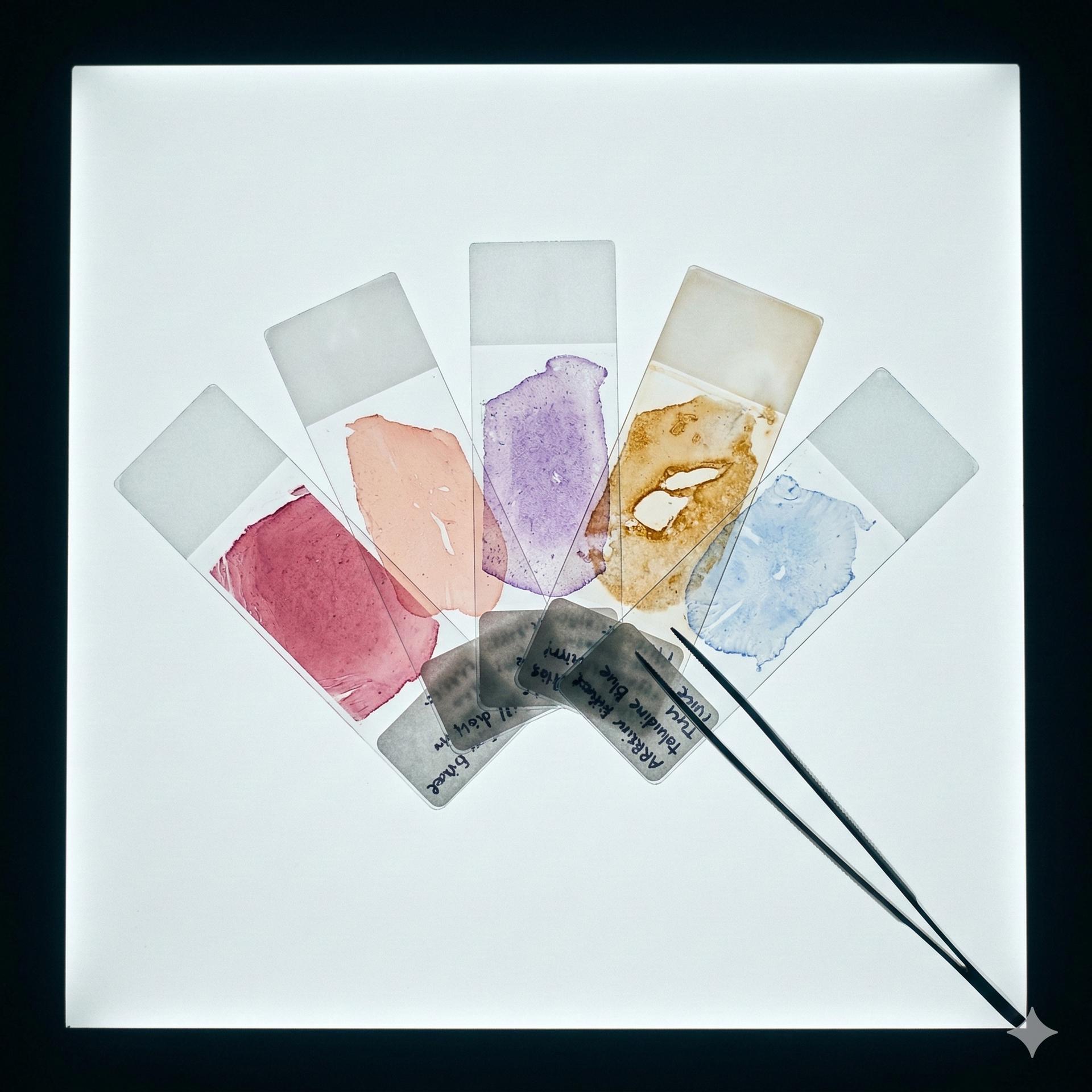
Mohs Lab: Tissue Processing & Frozen Section Histology
The Mohs laboratory is the backbone of Mohs micrographic surgery. Every step, from tissue accessioning through frozen section interpretation, directly determines cure rates. A single processing error can obscure residual tumor and lead to incomplete excision. This article covers the complete Mohs lab workflow: specimen handling, tissue grossing and mapping, cryomold embedding, cryostat microtomy, H&E and toluidine blue staining protocols, immunohistochemistry for melanoma and perineural invasion, and quality assurance standards. Practical troubleshooting tables address the most common staining artifacts encountered in daily practice.
CPT Coding & Billing for Mohs Surgery
Correct CPT coding ensures appropriate reimbursement, legal compliance, and accurate documentation of surgical complexity. This article covers the complete coding framework for Mohs micrographic surgery (17311–17315), skin biopsies (11102–11107), destruction of premalignant and malignant lesions (17000–17286), excisions (11400–11646), shave removals (11300–11313), wound repair hierarchy (simple, intermediate, complex), adjacent tissue transfers (14000–14302), interpolation flaps, skin grafts (15100–15261), and cartilage grafts. Key modifiers (-25, -51, -58, -59, -76, -78, -79) are explained with clinical scenarios. CMS documentation requirements for Mohs surgery, CLIA laboratory regulations, OSHA workplace safety, and incident-to billing rules are summarized.
Anatomy
Danger zones, motor nerve pathways, undermining planes, and tissue layers.
Surgical Danger Zones
Critical neurovascular structures at risk during cutaneous surgery. Covers the three major motor nerve danger zones (spinal accessory, temporal branch, marginal mandibular branch), sensory nerve anatomy including the trigeminal and cervical plexus, and clinically important syndromes resulting from nerve injury.
Undermining Planes by Anatomical Region
Detailed guide to safe undermining planes for every region of the face, scalp, and neck. Correct plane selection is essential for tissue mobilization while avoiding injury to critical neurovascular structures. Each region has a specific safe plane dictated by the depth of vital structures.
Instruments
Surgical instruments, setup, and equipment for Mohs and reconstruction.
Regional Reconstruction
Region-specific reconstruction algorithms for nose, ear, eyelid, lip, and more.
Nasal Reconstruction
The nose is the most frequently reconstructed site after Mohs surgery. Nasal reconstruction demands respect for the subunit principle (Burget & Menick), three-layer anatomy (lining, framework, cover), and a systematic algorithmic approach based on defect size, depth, and subunit location. Options range from primary closure and bilobed flaps for small defects to paramedian forehead flaps for large or full-thickness defects.
Ear Reconstruction
Ear reconstruction after Mohs surgery presents unique challenges due to the thin skin with minimal subcutaneous tissue, complex three-dimensional cartilaginous framework, and the dual surface nature of the auricle. The reconstructive ladder ranges from secondary intention healing for concave surfaces to wedge excision, Antia-Buch advancement, retroauricular interpolation flaps, revolving door flaps, composite grafts, and skin grafts. Preserving helical rim contour and avoiding ear canal stenosis are paramount functional goals.
Eyelid Reconstruction
Eyelid reconstruction after Mohs surgery demands precise understanding of lamellar anatomy (anterior lamella: skin + orbicularis oculi; posterior lamella: tarsus + conjunctiva) and adherence to the cardinal rule that both lamellae must never be grafted simultaneously. The reconstructive algorithm is driven by the percentage of eyelid margin loss, with techniques ranging from direct closure for small defects to Tenzel semicircular rotation, Hughes tarsoconjunctival flap, Cutler-Beard bridge flap, and Mustarde cheek rotation for progressively larger defects. Ectropion prevention is a primary functional goal.
Lip Reconstruction
Lip reconstruction after Mohs surgery requires meticulous attention to key landmarks (vermilion border, wet line, Cupid's bow, philtral columns, oral commissures) and the primary functional goal of oral competence. The reconstructive algorithm is driven by the percentage of lip width lost: primary closure for defects under one-third, Abbe cross-lip flap for one-third to two-thirds, and Karapandzic or Bernard-Burow techniques for defects exceeding two-thirds. The double V-Y island pedicle technique addresses defects crossing the vermilion border.
Cheek, Temple, Forehead & Scalp Reconstruction
The cheek, temple, forehead, and scalp represent distinct reconstructive zones with varying tissue characteristics and anatomic hazards. The cheek offers generous tissue laxity favorable for local flaps and primary closure. The temple is a danger zone requiring superficial undermining to protect the temporal branch of CN VII. The forehead allows primary closure along RSTLs and A-T/O-T plasty for circular defects. The scalp has limited elasticity, requiring rotation flaps, galeal scoring, and subgaleal undermining for larger defects.
Tumor Types & Indications
Skin cancer types, mimickers, and precursor lesions. BCC, SCC, melanoma, MCC, DFSP, sebaceous CA, MAC, EMPD, AFX/PDS, and more. NCCN-based indications and treatment algorithms.
Basal Cell Carcinoma: Mohs Indications & Treatment
Basal cell carcinoma (BCC) is the most common malignancy in humans, with over 4 million cases diagnosed annually in the United States alone. While BCC rarely metastasizes, locally aggressive subtypes can cause significant tissue destruction if inadequately treated. This article reviews BCC histopathologic subtypes, NCCN v1.2026 risk stratification criteria, indications for Mohs micrographic surgery, AUC guidelines, and the treatment algorithm from low-risk to advanced disease.
Squamous Cell Carcinoma: Mohs Indications & Treatment
Cutaneous squamous cell carcinoma (cSCC) is the second most common skin cancer and carries a meaningful risk of local recurrence, perineural invasion, and regional/distant metastasis. Unlike BCC, high-risk SCC can be lethal. This article covers histopathologic subtypes and differentiation grading, NCCN v1.2026 risk stratification, the BWH T-staging system, indications for Mohs micrographic surgery, sentinel lymph node biopsy considerations, and emerging immunotherapy options for advanced disease.
Melanoma & Mohs Surgery: Indications and Slow Mohs Technique
Melanoma is the deadliest form of skin cancer, responsible for the majority of skin cancer deaths despite representing only 4-5% of skin cancer diagnoses. While standard wide local excision with predetermined margins remains the mainstay of surgical treatment, Mohs micrographic surgery, particularly the "slow Mohs" technique with immunohistochemistry. Plays an increasingly important role for melanoma in situ and lentigo maligna on cosmetically sensitive facial sites. This article reviews melanoma subtypes, NCCN v1.2026 margin guidelines, the slow Mohs technique, SLNB criteria, and modern adjuvant therapy options.
Merkel Cell Carcinoma: Mohs Surgery & NCCN Management
Merkel cell carcinoma (MCC) is a rare, aggressive neuroendocrine skin cancer with rising incidence. Associated with Merkel cell polyomavirus (MCPyV) in approximately 80% of cases, MCC primarily affects older and immunosuppressed patients on sun-exposed sites. NCCN v2.2026 guidelines recognize Mohs micrographic surgery (PDEMA) as an appropriate surgical approach, alongside wide excision. This article covers epidemiology, clinical presentation, AJCC staging, the complete NCCN treatment algorithm including neoadjuvant immunotherapy, the specific role and considerations of Mohs in MCC, systemic therapy for advanced disease, and surveillance recommendations.
DFSP: Mohs as Treatment of Choice (NCCN v2.2026)
Dermatofibrosarcoma protuberans (DFSP) is a rare, locally aggressive fibroblastic tumor with low metastatic potential but extensive subclinical extension. NCCN v2.2026 recommends Mohs micrographic surgery (PDEMA) as the preferred surgical approach due to superior local recurrence rates compared to wide excision. This article covers the pathognomonic COL1A1::PDGFB translocation, CD34+ immunophenotype, NCCN treatment algorithm, the critical importance of delayed reconstruction, fibrosarcomatous transformation (FS-DFSP), and imatinib for unresectable or metastatic disease.
AFX & Pleomorphic Dermal Sarcoma: Mohs Indications & Treatment
Atypical fibroxanthoma (AFX) and pleomorphic dermal sarcoma (PDS) represent a spectrum of UV-induced cutaneous sarcomas arising on sun-damaged skin of elderly patients. AFX is confined to the dermis with excellent prognosis (<1% metastasis), while PDS extends into the subcutis and/or harbors adverse histologic features (necrosis, lymphovascular invasion, perineural invasion) carrying 10-20% metastatic potential. Both are diagnoses of exclusion. The hallmark IHC finding is negativity for all lineage-specific markers (cytokeratins, S-100, desmin, Melan-A), requiring systematic exclusion of spindle cell SCC, melanoma, leiomyosarcoma, and angiosarcoma. Mohs micrographic surgery is the treatment of choice for both AFX and PDS, offering superior margin control and local recurrence rates. For PDS with high-risk features, adjuvant radiation therapy and sentinel lymph node biopsy should be considered. This article provides a thorough overview of the AFX-PDS spectrum with emphasis on histopathologic diagnosis, IHC workup, Mohs surgical technique, and management algorithms.
Sebaceous Carcinoma: Mohs Indications & Treatment
Sebaceous carcinoma (SC) is a rare, aggressive adnexal malignancy arising from sebaceous glands, most commonly in the periocular region where it accounts for approximately 1-5% of eyelid malignancies. Notorious for its clinical mimicry of benign conditions such as chalazion and chronic blepharoconjunctivitis, SC is frequently diagnosed late, contributing to significant morbidity and mortality. Pagetoid intraepithelial spread is a hallmark feature that complicates margin assessment and necessitates specialized techniques including map biopsies and immunohistochemical staining on Mohs frozen sections. Association with Muir-Torre syndrome (mismatch repair deficiency) mandates screening for internal malignancies in select patients. This article reviews epidemiology, clinical presentation, histopathology, immunohistochemistry, staging, the role of Mohs micrographic surgery with immunostain-guided margin assessment, treatment algorithms including sentinel lymph node biopsy and adjuvant therapy, and prognosis.
Extramammary Paget Disease (EMPD): Mohs Indications & Treatment
Extramammary Paget disease (EMPD) is a rare cutaneous adenocarcinoma arising in apocrine gland-bearing skin, most commonly the vulva, perianal region, groin, axillae, and penile/scrotal skin. Clinically mimicking chronic eczema or dermatitis, EMPD is frequently misdiagnosed for years before biopsy. The critical distinction between primary EMPD (in situ apocrine adenocarcinoma, CK7+/CK20-) and secondary EMPD (cutaneous extension of an underlying visceral malignancy, often CK20+) has profound implications for workup and prognosis. Mohs micrographic surgery is an excellent indication for EMPD due to the hallmark extensive subclinical extension with irregular margins that routinely extend 2-5 cm beyond the visible clinical border. Immunostain-guided Mohs (CK7 on frozen or permanent sections) dramatically improves margin assessment compared to standard H&E. Wide local excision carries recurrence rates of 30-60%, while Mohs reduces recurrence to 8-16%. This article covers epidemiology, clinical presentation, histopathology and IHC profiling, primary versus secondary EMPD differentiation, Mohs surgical technique, staging of invasive disease, non-surgical therapies, and prognosis.
Microcystic Adnexal Carcinoma (MAC): Mohs Indications & Treatment
Microcystic adnexal carcinoma (MAC) is a rare, locally aggressive appendageal neoplasm with dual follicular and eccrine differentiation. MAC has a striking predilection for the centrofacial region, particularly the nasolabial fold and upper lip. And is characterized by extensive subclinical extension, dense sclerotic stroma, and a remarkably high rate of perineural invasion (~80%). Despite its locally destructive behavior, distant metastasis is exceedingly rare (<1%). Mohs micrographic surgery is the gold standard treatment, with recurrence rates under 5% compared to 40-60% with standard wide local excision. This article covers the epidemiology, clinical presentation, characteristic histopathology with dual differentiation, critical differential diagnosis (desmoplastic trichoepithelioma, morpheaform BCC, syringoma), Mohs surgical technique including the role of slow Mohs, perineural invasion management, the treatment algorithm, and long-term prognosis.
Dysplastic Nevus: Surgical Indications & Melanoma Risk
Dysplastic nevi (atypical melanocytic nevi, Clark nevi) are melanocytic proliferations that occupy a morphologic and biologic continuum between common acquired nevi and melanoma. Present in 2-10% of the general population, they serve as both independent markers of elevated melanoma risk and potential direct precursors to melanoma. The clinical significance of a dysplastic nevus depends on the grade of histologic atypia (mild, moderate, or severe), margin status on biopsy, the total nevus count, family history of melanoma, and the presence of Familial Atypical Multiple Mole and Melanoma (FAMM) syndrome. While Mohs micrographic surgery is not the standard treatment for dysplastic nevi, understanding their management is essential for the Mohs surgeon who frequently encounters them during skin cancer screening and must make re-excision decisions. This article reviews the histopathologic grading, surgical management algorithm by atypia grade, FAMM syndrome, the molecular pathway from nevus to melanoma, and evidence-based surveillance protocols.
Genital Intraepithelial Neoplasia & SCC In Situ: Mohs Indications
Penile and genital intraepithelial neoplasia (PeIN) encompasses a spectrum of premalignant and in situ squamous neoplasms of the genital skin including Bowen disease, erythroplasia of Queyrat, and bowenoid papulosis. The WHO 2016 classification divides PeIN into undifferentiated (HPV-driven, p16-positive) and differentiated (lichen sclerosus-associated, p53-aberrant) subtypes, with the latter carrying a higher risk of progression to aggressive invasive SCC. Mohs micrographic surgery is a critical treatment modality for genital SCC in situ given the paramount importance of tissue conservation in the genital region, the high rate of subclinical extension, and elevated recurrence rates with standard excision. This article covers the complete classification system, clinical presentation of each entity, histopathologic and immunohistochemical features, risk of progression to invasive disease, surgical and non-surgical treatment approaches, management of invasive genital SCC, and prognosis with follow-up recommendations.
BCC & SCC Mimickers: Histologic Pitfalls for Mohs Surgeons
Accurate intraoperative histologic interpretation is the cornerstone of Mohs micrographic surgery. Benign adnexal tumors (trichoepithelioma, trichoblastoma, basaloid follicular hamartoma) can closely mimic basal cell carcinoma, while reactive proliferations (pseudoepitheliomatous hyperplasia, irritated seborrheic keratosis) and keratoacanthoma can be indistinguishable from squamous cell carcinoma on frozen sections. Misinterpretation in either direction carries significant consequences: overcalling a benign mimicker as carcinoma leads to unnecessary tissue removal, while undercalling true carcinoma leaves residual tumor. This article provides a systematic approach to the most common BCC and SCC mimickers encountered during Mohs surgery, with detailed immunohistochemical panels, comparison tables, and practical decision algorithms for the Mohs surgeon.
Systemic Therapy & Radiation for Cutaneous Malignancies
A growing subset of cutaneous malignancies exceeds the reach of surgery alone. This article reviews FDA-approved systemic therapies and radiation indications across the five major cutaneous malignancies encountered by Mohs surgeons: BCC (hedgehog pathway inhibitors, cemiplimab), CSCC (cemiplimab, pembrolizumab), MCC (avelumab, pembrolizumab), melanoma (PD-1 inhibitors, BRAF/MEK inhibitors, ipilimumab), and DFSP (imatinib). For each agent, mechanism of action, dosing, response rates, key adverse effects, and monitoring requirements are detailed. A dedicated section addresses radiation therapy indications including definitive, adjuvant, and salvage settings. The article also covers immune-related adverse event recognition and graded management, with particular attention to dermatologic irAEs relevant to the Mohs surgeon.
Tumor Biology & Molecular Pathways in Cutaneous Oncology
Understanding the molecular pathogenesis of cutaneous malignancies is foundational for Mohs surgeons. Each major skin cancer type is driven by distinct genetic alterations: BCC by Hedgehog pathway mutations (PTCH1, SMO), SCC by TP53 and NOTCH inactivation, melanoma by MAPK pathway activation (BRAF, NRAS), MCC by Merkel cell polyomavirus T antigen oncoproteins or UV-induced mutations, and DFSP by the COL1A1-PDGFB fusion translocation. Beyond sporadic tumors, hereditary cancer syndromes such as Gorlin syndrome, xeroderma pigmentosum, Li-Fraumeni syndrome, and Muir-Torre syndrome predispose patients to multiple cutaneous malignancies requiring lifelong surveillance and frequently repeated Mohs procedures. This article reviews the key molecular pathways, actionable mutations, and genetic syndromes relevant to dermatologic surgery practice.
Primary Cutaneous Mucinous Carcinoma
Primary cutaneous mucinous carcinoma (PCMC) is a rare low-grade adnexal carcinoma characterized by tumor cells floating in pools of extracellular mucin. It most commonly presents as a slow-growing pink-erythematous nodule on the periorbital region or head and neck in older adults. Histologically, the defining feature is nests of epithelial cells within lakes of mucinous material separated by fibrovascular septa. Immunohistochemistry (CK7+, CK20−) distinguishes PCMC from metastatic mucinous adenocarcinoma of gastrointestinal origin (CK20+); synaptophysin and chromogranin positivity is seen in the neuroendocrine subtype only. Endocrine mucin-producing sweat gland carcinoma (EMPSGC) is recognized as the in situ precursor of neuroendocrine-type PCMC. Treatment involves surgical excision with margin control, and Mohs micrographic surgery is particularly advantageous for periorbital tumors where tissue conservation is critical. Overall prognosis is favorable, with a distant metastasis rate of approximately 5–6% and a 9% risk of subsequent primary malignancy at another site.